The contents are the result of pregnancies that ended in the early stages and the tissues were washed to remove blood and menstrual residues. Look at the pictures!
You may have already seen on the Internet alleged images of what a baby would look like in early pregnancy, especially in sensationalist materials against the decriminalization of abortion. The problem, however, is that these images showing the shape of a child do not correspond to reality, since up to the 9th week the gestational material is still at the beginning of development and does not have defined contours.
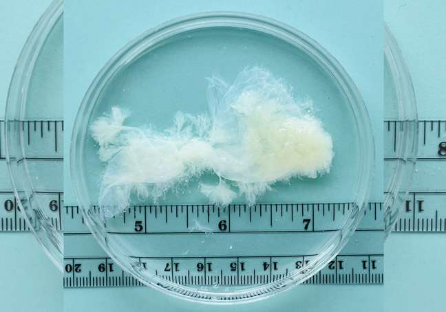
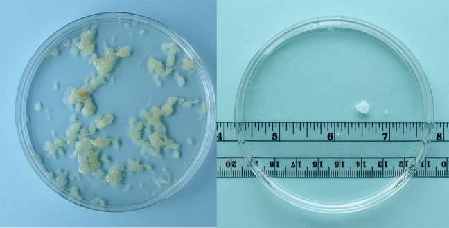
To clarify this information, the MYA Network in the United States started the project The question of the fabric (in literal translation, “the matter of fabric”), which is seen in very sharp photos what is eliminated from the uterus in an abortion performed in the first few weeks.
The intention in spreading these images is to fight the disinformation circulating on the networks and in common sense, leading women to a real knowledge of the functioning of their own body. The contents shown in the Petri dishes (those placed under the microscope) are the result of early pregnancy interrupted, whose tissues were removed by manual suction, remaining almost intact. Then they were washed to remove the blood and menstrual lining.
The project, in addition to spreading the images, also explained the sound of the so-called heartbeats which can be heard as early as the sixth week of pregnancy. According to Joan Fleischman, physician, researcher and activist who founded the MYA network, there is not yet a formed heart; what happens is that the cells that will unite to give rise to the organ already exist and will “beat” together – here’s where the sound you hear on the ultrasound.

“I was motivated by my patients to create this project”, says Fleischman in the project’s ad video. “Sometimes, they have chosen to see their gestational tissue in my office and, when they saw it, they were relieved, surprised or even intrigued that it looked nothing like what they had seen on the Internet. Seeing these reactions, I realized it was important to make these images publicly available. I think they speak for themselves. “
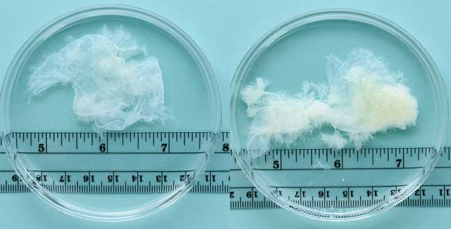
According to information released by the project, the gestational sac grows, on average, by 1 millimeter per day. Experts also explain that if a woman has an involuntary miscarriage or causes termination of pregnancy by taking abortion pills, she is likely to see a heavy menstrual flowwhich may be accompanied by blood clots of various sizes – different from the photos, which only show the gestational sac. “(…) so it can be difficult to see the pregnancy tissue, unless you look for it on purpose. If you’ve been pregnant for more than nine weeks and choose to look, you can see an early embryo,” they warn.
Watch the video where Joan Fleischman presents the project below. If you prefer, click on gear icon> subtitles> automatic translation to follow in Portuguese.
+The best content in your email for free. Choose your favorite Earth Newsletter. Click here!
Source: Terra
Benjamin Smith is a fashion journalist and author at Gossipify, known for his coverage of the latest fashion trends and industry insights. He writes about clothing, shoes, accessories, and runway shows, providing in-depth analysis and unique perspectives. He’s respected for his ability to spot emerging designers and trends, and for providing practical fashion advice to readers.


