With a technique that uses special ink and ultrasonic waves, scientists are able to solidify materials inside the human body, in a non-invasive 3D print
An innovative technique will allow doctors to make 3D prints with waves ultrasound inside the human body, creating organ parts and structures and delivering drugs non-invasively. More efficient than the previous method, which involved light, the new technology uses sound-sensitive injected inks and is called Deep Penetration Volumetric Acoustic Printing (DVAP).
To the ultrasonic waves they penetrate more than 100 times deeper into the tissues of the human body, while still maintaining spatial precision. This is thanks to the sonothermal effect, when sound waves are absorbed and increase the temperature. This solidifies the ink placed in the patient and creates the structures needed to perform 3D printing.
How to print with ultrasound
The basis of the technique is a special type of ink called sonic dye, composed of hydrogels, microparticles and water-reactive molecules. ultrasonic waves. The viscous liquid is easily injected into targets on the body, and through an ultrasonic printing probe, waves are projected into the ink, causing its particles to stick together and solidify into complex structures, ranging from scaffolds that mimic bones to hydrogel bubbles. used for adaptation of the organ.
")
After the procedure, the remaining paint can be removed with a syringe. The substance is versatile and its formula can be modified to have more or less durability, degradation capacity and even different colors. The demonstration of the potential of DVAP technology was carried out through three different tests.
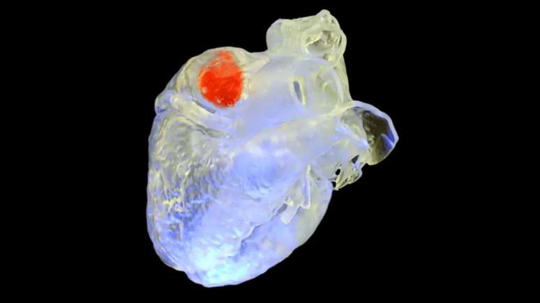
In the first test, sonic dye was used to seal a section of a goat’s heart, which typically requires open-chest surgery. Penetrating through 12 mm of tissue, the procedure glued the paint to the heart tissue without causing damage, and the flexible material successfully supported the animal’s heartbeat.
In the second test, the reconstruction and regeneration capabilities were demonstrated by injecting the substance into a defective chicken leg, attaching to the bone through 10 mm of skin and muscle without damaging surrounding tissue. In the latest test, a common chemotherapy drug was injected into the liver, where solidified hydrogels slowly released the drug.
Although it is far from reaching hospitals, the technique promises great potential in surgeries and therapies that would normally be quite invasive and destructive to the human body.
Source: Science, Duke Pratt School of Engineering
Trends on Canaltech:
- What’s happening with Xiaomi?
- Netflix not working? Streaming is down and users are complaining
- Charlie and the Chocolate Factory | What’s the cast like these days?
- The 50 funniest Google Assistant jokes
- Apple releases iOS 17.2 with Journal app and camera improvements
Source: Terra
Rose James is a Gossipify movie and series reviewer known for her in-depth analysis and unique perspective on the latest releases. With a background in film studies, she provides engaging and informative reviews, and keeps readers up to date with industry trends and emerging talents.


 and ventured into Cinemaporpor")

