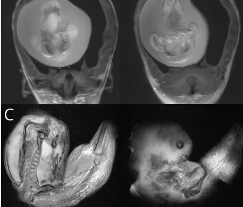
Surgery to remove a “parasitic twin” with spine, femur and tibia took place in China and was reported in a scientific paper. Understand!
A report published by neurology.org has attracted the attention of the medical community: in China, a one-year-old girl had a fetus removed from her brain. The situation is unusual and rare, but it is present in the scientific literature as what it is called fetus-in-fetu or “parasitic twin”.
The most accredited hypothesis for this type of case is that, during pregnancy, which initially would be twins, the cells of a malformed embryo are incorporated by the healthy embryo. Thus, both continue to grow until the one that has been incorporated stops at a certain point (although it may continue to grow if it receives blood flow).
The story was reported by Chunde Li, of Beijing Tiantan Hospital, in the Chinese capital. Second Medpage Todayabout 200 cases of parasitic twins have been documented in the medical literature (the first in 1808), but very few of them were located in the skull.
It is important to underline this it is not a teratoma, it is a tumor of embryonic origin formed by tissues (such as teeth, hair and muscles), usually found in the ovaries or testicles. According to Chunde Li, what differentiates the fetus-in-fetu is the presence of vertebrae and internal organs, in addition to the fact that it is found in very young patients.

Removing the parasitic twin
According to the article, the girl had delays in motor development – could not, for example, remain seated without support. Also, the skull circumference was greater than expected, although there were no signs of intracranial hypertension, such as nausea, vomiting, or eye changes.
During the investigative investigations it was discovered, through computed tomography and magnetic resonance, that the girl had hydrocephalus, cerebral compression and a fetiform mass about 10 cm long, showing vertebral column, femur and tibia. The images revealed that the parasitic twin had spina bifida, and on further examination, the upper limbs and finger-like structures were also noted.
to access the full article. The report does not provide information about the girl’s postoperative period.
Source: Terra
Ben Stock is a lifestyle journalist and author at Gossipify. He writes about topics such as health, wellness, travel, food and home decor. He provides practical advice and inspiration to improve well-being, keeps readers up to date with latest lifestyle news and trends, known for his engaging writing style, in-depth analysis and unique perspectives.


